As the child of two veterinarians in practice, one of who became a research scientist, you could say I grew up around medicine and science.We lived in northern Sydney, in a house that was also the Veterinary Hospital, sandwiched between the railroad tracks out back and the Pacific Highway in the front. In the operating theatre and examination room, I watched birth and death, and operations being performed out of books, while I ‘assisted’ by opening sutures and powdering gloves from the early age of five.
Fifteen years later, as an undergraduate in photography, I asked my father (by that time a veterinary pathologist and research scientist at Cornell University in upstate New York), to take a muscle biopsy from me so that I could make a microscopic self-portrait. He balked at the idea, citing it would be painful and unnecessary, so instead I loitered around the post-mortem area at Cornell. Here, dismembered horse legs stuck out of 44-gallon barrels on the landing and you donned galoshes to wade through the organic muck on the floor. My twin-lens reflex camera was flecked with bits of cow insides as I made tableaux style photographs. The dramatic setting of stainless steel, fluorescent lighting, blood and guts fascinated me – all more ‘Film Noir’ than small town. Later I would use animal tissue supplied by dad to make a series of images using light microscopes.
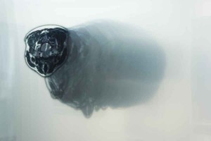
All of this was a prelude to my first major art–science collaboration, RAPT I and II, now celebrating their ten-year anniversary. At Sydney College of the Arts, I devised a project for my Masters that spoke about modern technology’s ability to form and change our conception of space and time. Such an undertaking required some sort of grounding and focus. I decided on using the body in combination with medical imaging technology as a metaphor. First, I needed my full body MRI scanned. Second, I needed a way of translating the information into something I could use. And finally, I needed a place with sufficient processing power and the software necessary to manipulate the data set. While I don’t have the methodology of a scientist, this first major project was a fundamental lesson for me in grasping that most art and science is procedural, with potential outcomes that can transcend the process and have greater meaning.
In practical terms, all collaborations begin with people, not institutions. For RAPT, I was introduced to a doctor who worked with a private imaging lab. The lab had an MRI technologist who was willing to come in out of hours and perform the six hours of imaging needed to produce a full data set. She knew someone at Westmead Children’s Hospital who could translate the proprietary format of the scans into generic TIFF files. My thesis supervisor, Geoff Weary, knew the Director of VisLab – a high-performance visualisation laboratory, at the time located in the Physics Department at Sydney University – and was setting up the possibility of students working there. I took my data set to VisLab and used their computational power and their licensed software, Voxelview® and VoxelAnimator®, for volume rendering and animating. Though the render farms of today dwarf the Cray processor at the old VisLab, without access to the state-of-the-art (for the time) facilities and abilities of each place and person, RAPT would not have manifested. For the contributors, it was a refreshing take on their own medium. While it was an artwork, it was also a catalyst for me to continue making work that crossed boundaries and disciplines.
A year later (1999), with a new media grant from the Australia Council, I began work on Scynescape. This project was formulated as a maze-like immersive environment using sensors, projectors, sound and latex. The content was generated from a scanning electron microscope (SEM), using as subjects moulds taken from the surface of my body and hair and then coated with gold to allow them to conduct a high-energy beam of electrons. This conductivity renders the surface topography of the specimen. I was hosted by the University of Sydney’s EMU (Electron Microscope Unit), part of the Australian Key Centre for Microscopy and Microanalysis. With considerable help from the lab technician in preparing the samples, and general training and guidance in the use of the electron microscope, including hacking it to go straight out to videotape, I was granted access. Usually I took the graveyard shift, when the machine was least in demand. However, the times when there were more scientists floating through were much more enriching. I remember a team coming by while I had a hair sample on the microscope stage. A discussion ensued about the physical nature of hair and whether it looked different or similar in different ethnicities. This would prove a useful bit of information to me in a later project, Transformers. Once again, the facility and access to equipment has its purpose, but the human interaction holds the possibility.
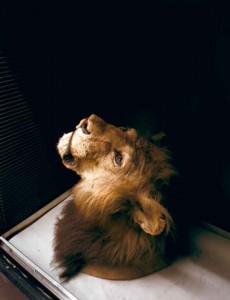
An ANAT Synapse residency at The American Museum of Natural History (New York) in 2002 to work on Transformers in their genetics lab jump-started a further residency that resulted in the Saved By Science series (2004–2005). Saved By Science marked a turning point in my work, in that I was not using the tools of science or medicine. Instead the project regarded the institution of science itself; specifically the collecting and ordering of the natural world in our quest to understand where we come from. To create a project which is inherently about the position of science in culture required a level of immersion only attainable by being on the inside. The unprecedented access granted was critical to the work, and the availability of over 200 researchers and scientists with whom to speak was invaluable.
The technologies of science progress – imaging technologies improve and DNA sequencing become more efficient. The technologies of media arts also shift and morph. Not so long ago, CD-ROMs created with Director® software were a preferred interactive platform, DVD authoring was cutting edge and projectors were prohibitively expensive. However, these are only technologies and without content they obviously lack meaning. What becomes lasting in collaborations and connections between different disciplines includes the exchange of thought, the glimpse into another modus operandi and the stretch required to bridge the gap. For me, it’s the elastic nature of that connecting – of going to places I may or may not think I understand, of conversing with individuals that look at a body of knowledge with a wholly different methodology – that has fed my creative inquiry and produced anything at all of interest.
Justine Cooper
Justine Cooper’s interdisciplinary artwork investigates the intersections between culture, science and medicine. She moves between various media, as well as medical imaging technologies such as MRI, DNA sequencing, ultrasound and scanning electron microscopy (SEM). Justine’s work has been recognised internationally and exhibited worldwide. She is currently working on a new series focused on medical simulation centres in Australia and the USA.
Read More
Watch More
 This work is licensed under a Creative Commons Attribution-NonCommercial-ShareAlike 2.5 Australia.





I have been looking looking around for this kind of information. Will you post some more in future? I’ll be grateful if you will.
Hi there, you might be interested in looking at our art science program sites including http://www.synapse.net.au and http://www.superhuman.org.au/